
Imagine if cells could attend an art class. Instead of brushes and canvases, they’d use fluorescent dyes and high-throughput microscopes to create their own masterpieces. Welcome to the world of Cell Painting, where biology meets art in the most colorful way possible! This innovative technique doesn’t just make pretty pictures; it reveals the hidden secrets of cellular life by highlighting key components and organelles.
Cell Painting is a multiplexed image-based and high-throughput phenotypic profiling assay that uses molecular “paints” to visualize and measure the organization of cellular features as read-outs of cellular state. Cell Painting provides comprehensive morphological data used to predict compound activities and potential hazards in drug discovery and toxicology.
The technique is valuable in both preclinical and early discovery programs as it helps discover disease mechanisms, infer new mechanistic insights, refine target identification, and uncover off-target effects and cytotoxicity.
Intrigued by Cell Painting and want to discover how it could help your drug discovery or development pipeline? Read on to find out more.
A short history of Cell Painting
The Cell Painting concept was introduced in 2013 by Gustafsdottir et al. to help researchers detect changes in cell states associated with different compound treatments or diseases. Initially, it was limited to detecting seven cell components using six stains: nucleus, endoplasmic reticulum, nucleoli, Golgi apparatus, plasma membrane, F-actin, and mitochondria.
Since then, the assay has been successfully implemented, optimized, and standardized at various locations, leading to several updates, such as improved stain quality, quantitative optimization, simplified steps, or cost-saving due to reduced stain concentrations (Bray et al., 2016; Cimini et al., 2023). In particular, the Broad Institute of Harvard and MIT pioneered the use of numerous fluorescent morphological markers in Cell Painting, ensuring the method’s scalability for large experiments (Bray et al., 2016).
A brief description of the general Cell Painting workflow
For the Cell Painting assay, cells are plated in multiwell plates—usually 384-well plates—and treated with the compounds of interest. They are then subsequently fixed (typically with paraformaldehyde), stained with six fluorescent dyes (Table 1) that label eight distinct sub-cellular compartments and organelles, and imaged using a high-throughput microscope (Fig. 1 and 2). Due to the limited number of available microscopic channels, some dyes, such as those for Actin and Golgi, are merged. When four channels are used, another two dyes, like those for RNA and ER, are combined into a single channel.
Table 1 – The list of dyes used in the Cell Painting assay
|
Dye |
Organelle or cellular component |
|
Hoechst 33342 |
Nucleus |
|
Concanavalin A/Alexa Fluor 488 conjugate |
Endoplasmic reticulum |
|
SYTO 14 nucleic acid stain |
Nucleoli, cytoplasmic RNA |
|
Phalloidin/Alexa Fluor 568 conjugate, WGA/Alexa Fluor 555 conjugate |
F-actin cytoskeleton, Golgi apparatus, plasma membrane |
|
MitoTracker Deep Red |
Mitochondria |
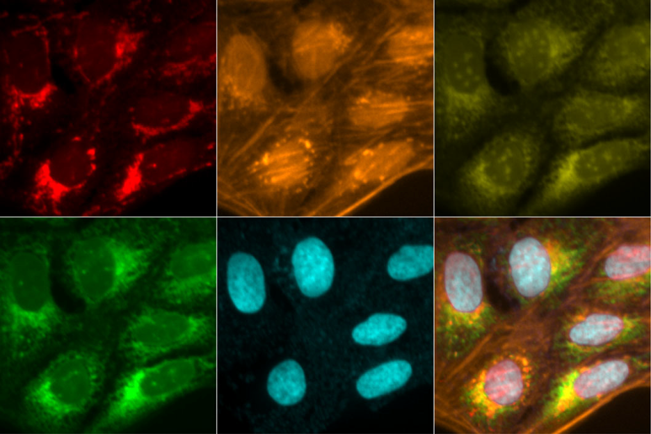
Figure 1- U2OS cells subjected to Cell Painting assay, using the six dyes in five channels to stain eight sub-cellular compartments and organelles. The top row (left to right): mitochondrial staining: actin, Golgi apparatus (red), and plasma membrane staining (orange), followed by nucleolar and cytoplasmatic RNA staining (yellow). The bottom row (left to right): endoplasmic reticulum staining (green), DNA staining (blue), and the overlap stains of all five channels(merge). Image taken from Cimini et al., 2023.
The 384-well plate is typically imaged at multiple positions in both horizontal (XY) and vertical (Z) dimensions to capture many cells and various sub-cellular regions. Consequently, the primary raw data from Cell Painting assays consist of thousands of multi-dimensional images at gigabyte-scale, with each image usually containing hundreds of cells.
During the subsequent image analysis step, these images are processed using automated tools integrated into proprietary software, like Harmony Revvity Inc., or open-source software such as CellProfiler (Bray et al., 2016; McQuin et al., 2018; Stirling et al., 2021). The software identifies individual cells and measures hundreds to thousands of morphological features, including size, shape, texture, and intensity, resulting in single-cell profiles. These features create morphological cell profiles that reflect the biological state of each cell and can detect subtle phenotypes. The Cell Painting workflow is represented in Fig. 2.
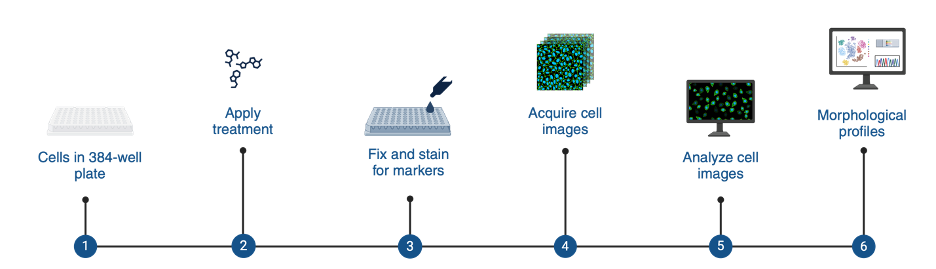
Figure 2- The Cell Painting workflow
Applications of Cell Painting
Individual cell profiles can be compared to achieve various objectives, such as identifying the effects of chemical or genetic perturbations based on their similar morphological effects, grouping compounds or genes into functional pathways, detecting disease signatures, and predicting assay outcomes using machine learning (Gustafsdottir et al., 2013). Consequently, Cell Painting is valuable in both preclinical and early discovery programs, as it offers the possibility to infer new mechanistic insights and promising alternatives to traditional binding assays typically used in training machine learning models.
Additionally, morphological cell features can potentially reveal the mechanisms of action of compounds in human cells. This information could be valuable in developing anti-tumor compounds or assessing the health and environmental risks of industrial chemicals (Nyffeler et al., 2023; von Coburg and Dunst, 2023).
In summary, the introduction of Cell Painting paves the way for a more comprehensive evaluation of compound effects. However, working with high-throughput cellular readouts presents significant challenges that must be addressed to fully leverage the data.
Possible limitations in Cell Painting assays
While it is undoubtedly a powerful method, Cell Painting assays have various limitations, including the types of cells suitable to investigate and computational difficulties in making reliable measurements or inferences.
For instance, the most commonly used cells for Cell Painting are the U2OS and A549 cell lines, given their relatively ‘generic’ type, ease of cultivation, and suitability for imaging as they are adherent, non-spheroid, and have limited clumping (Carpenter et al., 2016). Non-adherent cells are far less suited to this technique.
Also, different cell types might exhibit different phenotypic signatures when perturbed with diverse compounds, so selecting an appropriate cell line that is physiologically relevant to the area of study could require researchers to adjust protocols and image software analysis to maximize the likelihood of robust insights.
Additionally, given the limited number of features that can be identified using the Cell Painting assay, researchers typically have to computationally infer additional features, which might limit the discovery process and introduce biases. Any interpretations of morphological profiles in terms of underlying biology can also sometimes be non-trivial due to the high dimensionality of the data.
Alongside these issues, there are three main computational challenges faced.
Firstly, analyzing the high-dimensional feature space generated by Cell Painting involves statistical difficulties, such as spurious correlations and multiple testing.
Secondly, single-cell data requires significantly more computational storage and processing resources. Currently, there is no established routine analytical protocol to address this issue.
Lastly, analyzing and integrating data across different experiments is complex, necessitating proper control over the substantial effects of variations in cell seeding, growth, and other batch-related or systematic artifacts.
Similarly, while cell painting is high-content, processing large-scale compound libraries still requires substantial time and resources, however, integration with AI and deep learning methods stand to help overcome these challenges by providing automated feature extraction and predictive modeling, even at the single-cell level.
How does Cell Painting compare to other methods?
While various methods exist for generating rich profiles of biological samples, high-throughput transcriptomics technologies are currently the only practical alternative to Cell Painting in terms of throughput and efficiency. The Cell Painting assay offers single-cell resolution, whereas high-throughput gene-expression profiling methods, like L1000 or MERCURIUS™ DRUG-seq, characterize cell population-level responses.
Morphological profiling at the single-cell level can enhance the ability to detect changes in cell subpopulations, while high-throughput transcriptomics allows for screening more samples, cell types, drugs, and experimental conditions.
L1000 is a high-throughput bead-based gene expression profiling assay that directly measures the mRNA abundance of 978 “landmark” genes selected to represent the diversity of biological pathways and processes in human cells. Researchers use this subset of the transcriptome to computationally infer the expression of an additional 11,350 genes not explicitly measured (Subramanian et al., 2017). More details about the opportunities and challenges of the L1000 gene expression profiling technology can be found here.
MERCURIUS™ DRUG-seq is a transformative tool for compound screening and drug discovery, combining unbiased, high-throughput compound screening with massively parallel and extraction-free transcriptomics. This method uses highly optimized and rigorously evaluated sample barcodes and unique molecular identifiers to tag the 3’ poly(A) tail of all mRNA molecules in a sample-specific manner during the first-strand synthesis step of cDNA library preparation. The DRUG-seq protocol is designed to work with frozen cells and bypass RNA extraction. Thanks to the highly optimized lysis buffers for cell lysis of 2D cell cultures and organoid models, it efficiently generates library preps without prior RNA isolation.
Together, these methods are complementary, each providing unique insights that, when combined, can offer a more comprehensive understanding of cellular responses.
Conclusion
The Cell Painting assay holds great promise for predicting compound activities and hazards in drug discovery and toxicology. By combining Cell Painting-based phenotypic data with structural information from compounds, machine learning and deep learning models can predict compound activities across various disease endpoints and uncover mechanisms of action. This approach has the potential to enhance our understanding of compound responses within cells, guiding therapeutic development and risk assessment.
While Cell Painting offers significant potential for advancing compound evaluation and toxicology research, overcoming its challenges is essential to fully realizing its benefits. Collaborative efforts, standardization, and improved methodological approaches will enable the scientific community to harness the power of Cell Painting data for innovation and advancement.
References
- Bray, M. A., Singh, S., Han, H., Davis, C. T., Borgeson, B., Hartland, C., et al. (2016). Cell Painting, a high-content image-based assay for morphological profiling using multiplexed fluorescent dyes. Protoc.11, 1757–1774. doi:10.1038/nprot.2016.105
- Gustafsdottir, S. M., Ljosa, V., Sokolnicki, K. L., Wilson, J. A., Walpita, D., Kemp, M. M., et al. (2013). Multiplex cytological profiling assay to measure diverse cellular states. PLOS ONE8, e80999. Publisher: Public Library of Science. doi:10.1371/journal.pone.0080999
- Cimini, B. A., Chandrasekaran, S. N., Kost-Alimova, M., Miller, L., Goodale, A., Fritchman, B., et al. (2023). Optimizing the Cell Painting assay for image-based profiling. Protoc.18, 1981–2013. doi:10.1038/s41596-023-00840-9
- McQuin, C., Goodman, A., Chernyshev, V., Kamentsky, L., Cimini, B. A., Karhohs, K. W., et al. (2018). CellProfiler 3.0: next-generation image processing for biology.PLOS Biol. 16, e2005970. doi:10.1371/journal.pbio.2005970
- Stirling, D. R., Swain-Bowden, M. J., Lucas, A. M., Carpenter, A. E., Cimini, B. A., and Goodman, A. (2021). CellProfiler 4: improvements in speed, utility and usability.BMC Bioinforma. 22, 433. doi:10.1186/s12859-021-04344-9
- von Coburg, E., and Dunst, S. (2023). The adverse outcome pathway for breast cancer: a knowledge management framework bridging biomedicine and toxicology. Oncol. 14, 223. doi:10.1007/s12672-023-00840-x
- Subramanian, A. et al. (2017) ‘A Next Generation Connectivity Map: L1000 Platform and the First 1,000,000 Profiles’, Cell, 171(6), pp. 1437-1452.e17. Available at: https://doi.org/10.1016/j.cell.2017.10.049.
- Rohban, M. H. et al. Systematic morphological profiling of human gene and allele function via Cell Painting. Elife 6, e24060 (2017).
- Carpenter, A. E. et al. Cell Painting, a high-content image-based assay for morphological profiling using multiplexed fluorescent dyes. Nature Protocols, Vol,11, No. 9 (2016).