
Identifying brain-penetrant small-molecule modulators of human microglia using a cellular model of synaptic pruning – McCrea and Batorsky et al. [1]
Published May 9th, 2025, in Neuropsychopharmacology
Aims
- To screen 489 brain-penetrant small molecules with high-content imaging for the modulation of microglial synaptic pruning using a scalable, human-derived microglia model.
- To leverage MERCURIUS™ DRUG-seq to validate and enrich lead findings by uncovering diverse compound mechanisms of action affecting phagocytosis and inflammation.
- To prioritize potential therapeutic candidates for repurposing in neurological disorders.
Outcomes
- High-content imaging identified 28/489 compounds that significantly reduced microglial phagocytosis of synaptosomes by ≥50%.
- MERCURIUS™ DRUG-seq generated comprehensive transcriptomic profiles of these 28 hits and identified 16/28 “”RNA-active”” compounds, revealing diverse mechanisms suppressing phagocytosis and/or neuroinflammation.
- Integrated phenotypic and transcriptomic screening enabled the prioritization of therapeutics that modulate microglial function with minimal pro-inflammatory activation.
The Researchers
The study was led by researchers from the laboratory of Professor Roy H. Perlis, M.D, at the Center for Genomic Medicine and the Department of Psychiatry at Massachusetts General Hospital and Harvard Medical School, in collaboration with Dr. Rebecca E. Batorsky from the Data Intensive Studies Center at Tufts Institute for Artificial Intelligence, Tufts University, Medford, USA.
The Perlis lab uses cellular modeling, transcriptomics, clinical phenotyping, and small-molecule screening to develop novel therapeutics and clinical and genomic predictors of treatment response based on cellular models of brain disease, including schizophrenia, bipolar disorder, and depression.
The Challenge
Neurological disorders remain poorly treated due to a limited understanding of underlying mechanisms, a lack of robust biomarkers, and few functional therapeutic compounds. For instance, modulation of microglia is a promising therapeutic avenue as their dysregulation is implicated across a range of neurological disorders, including bipolar disorder, autism, and schizophrenia [2]. However, so far, screens have lacked scalable transcriptome-wide transcriptomic tools that provide robust mechanistic insights into drug activity at the gene expression level.
Traditional drug discovery pipelines rely on either high-throughput, target-based screening or phenotypic assays like high-content imaging techniques, such as Cell Painting [3]. While high-content imaging is scalable and excels at capturing morphological phenotypes, it fails to reveal transcriptional changes that result from treatment, hindering understanding of on- or off-target effects, toxic responses, or mechanisms of action [3].
Conversely, while traditional bulk RNA-seq approaches can reveal functional gene expression responses to potential therapeutic molecules, they largely lack the scalability for large compound libraries due to limited multiplexing capacity, expensive library preparation stages, and complex workflows [4]. Novel ultra-high-throughput 3’ bulk mRNA-seq approaches nowaddress this bottleneck by enabling low-cost transcriptome-wide profiling across thousands of samples [5].
In this study, the researchers aimed to uncover functional small-molecule modulators of microglial synaptic pruning [1]. Synaptic pruning by microglia is a core process in the human central nervous system, and its dysregulation is associated with the pathophysiology of many CNS disorders [2]. To address this, they combined large-scale high-content imaging with theultra-high-throughput transcriptomic technology, MERCURIUS™ DRUG-seq, to identify promising neuroactive compounds that generate diverse cellular, morphological, and transcriptomic responses.
Experimental Information
The team employed a high-content, scalable, and validated in vitro assay for microglia-mediated synaptic pruning using direct reprogramming of adult human peripheral mononuclear blood cells (PBMC) to induced microglia-like cells (piMGLCs) [6].
They then combined high-content phagocytosis assays with MERCURIUS™ DRUG-seq, enabling transcriptome-wide profiling of compound effects in a high-throughput format to refine their compound candidates and uncover diverse mechanisms of action.
Step 1: High-Content Functional Screening
- PBMCs were reprogrammed into piMGLCs using cytokine induction and displayed typical microglial markers and morphology after 10 days (Fig. 1).
- 489 CNS-penetrant small molecules were screened in a 96-well format by treating piMGLCs with 10 μM or 2 μM of compound for 24 hours (Fig. 1).
- Synaptosome phagocytosis was measured using pHrodo-labeled human synaptosomes imaged via confocal microscopy [7].
- Results from the initial screen were validated with an additional independent screen.
- Morphological profiling was performed on the validated hit list to inform on cellular phenotypes.
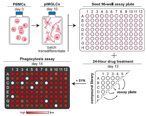
Figure 1. Workflow for deriving a large-scale batch of piMGLCs from PBMCs and subsequent functional synaptosome phagocytosis image-based screening in 96-well plates (Figure from McCrea and Batorsky et al., 2025).
Step 2: Transcriptomic Profiling with MERCURIUS™ DRUG-seq
- The refined compound hits confirmed in high-content and morphological profiling were assessed using Alithea’s MERCURIUS™ DRUG-seq kit.
- Sequencing was performed on an Illumina NovaSeq X Plus with a 1.5B flow cell.
- The researchers performed differential expression analyses, grouping of replicate wells using Uniform Manifold Approximation and Projection (UMAP) to demonstrate reproducibility of the treatment effects across wells and replicates, and functional enrichment analysis with QIAGEN Ingenuity Pathway Analysis (IPA).
Want to explore the robustness of MERCURIUS™ DRUG-seq data for yourself? Download our demo datasets for Hap1, HepG2, and HeLa cells to see the potential of high-throughput transcriptomics for your next screening experiment.
The Outcomes
High-Content Imaging Identifies Functional Hits
The high-content synaptosome phagocytosis assay on piMGLCs identified 47/489 compounds that showed ≥2 SD reduction in phagocytic index vs. control. The researchers confirmed the results from this primary screen with an independent experiment on the 47 hit compounds.
They then validated 28/47 compounds that reduced synaptosome phagocytosis by ≥50%. Compounds included kinase inhibitors, immunosuppressants, antipsychotics, and epigenetic modulators.
Morphological Profiling Provides Phenotypic Resolution
Microglia have exceptionally diverse morphological characteristics and actively modify the shape of their processes and soma in response to different stimuli. Here, the researchers used the degree of ramified (resting) vs. ameboid (active) morphotypes to indicate activation state [8].
Morphotypes varied across the 28 compound treatments. For instance, vorinostat induced hyper-ramified morphology (suggestive of suppressed activation), and cyclosporin A and lazertinib led to compact, ameboid forms (suggestive of an activated phenotype) (Fig. 2).
The different effects on morphology observed in response to compound treatments suggested they employed different molecular mechanisms to decrease phagocytic activity that could only be determined with transcriptomics.
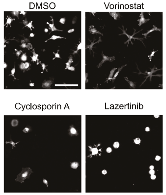
Figure 2. Phenotypic screening identified compounds that modulate piMGLC morphology, resulting in ramified (top row) or ameboid (bottom row) characteristics. Figure modified from McCrea and Batorsky et al., 2025.
High-Throughput Transcriptomics Adds Deep Mechanistic Insight
To decipher the cellular transcriptional mechanisms by which hit compounds affected microglial function, the researchers used MERCURIUS™ DRUG-seq to perform unbiased transcriptome-wide profiling across all 28 phenotypically relevant compounds following 24-hour treatment of piMGLCs.
Step 1: Discovering “RNA-Active” Compounds
Differential expression analysis found that 16 of the 28 hit compounds triggered significant changes in gene expression compared to DMSO controls (Fig. 3). The researchers termed these compounds “”RNA-active”.
The most transcriptionally active compound was the epigenetic modulator, vorinostat, which inhibits histone deacetylase (HDAC) and altered the expression of 3,525 genes relative to control (Fig. 3).
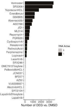
Figure 3. Secondary screening results with MERCURIUS™ DRUG-Seq identified differentially expressed genes (DEG) when comparing compound-treated samples to DMSO controls. Compounds with more than 95% DEGs of the DMSO vs. DMSO comparisons were termed “RNA-active”. Figure from McCrea and Batorsky et al., 2025.
Step 2: Uncovering High Reproducibility
The researchers then visualized the MERCURIUS™ DRUG-seq data from their 16 “RNA-active” hits with Uniform Manifold Approximation and Projection (UMAP) to assess reproducibility between replicates and visualize differences in responses for each compound.
Crucially, they found that treatment effects were highly reproducible across replicate wells and batches. A tighter grouping of replicates was observed for compounds that had a stronger impact on transcription, with clear distinctions in gene expression between different small molecules (Fig. 4).
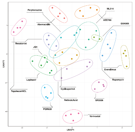
Figure 4. UMAP projection of the 16 active RNA hits shows grouping of replicates and clear transcriptional differences between compounds. Figure from McCrea and Batorsky et al., 2025.
Step 3: Revealing Diverse Mechanisms of Action
To reveal the mechanistic effects of different compound treatments, the researchers performed functional enrichment analysis on their MERCURIUS™ DRUG-seq data with QIAGEN’s Ingenuity Pathway Analysis (IPA). Functional enrichment analysis is a powerful method to detect dysregulated gene expression pathways that can help to explain phenotypes or morphologies discovered with high-content imaging.
Most compounds affected gene expression pathways involved in suppressing Fc gamma-receptor-dependent phagocytosis and phagosome formation (Fig. 5). This supported the initial high-content imaging experiments that screened for hits reducing synaptosome phagocytosis.
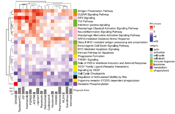
Figure 5. Transcriptomic pathway analysis uncovered diverse functional treatment responses. The heatmap (top) indicates the activation/suppression z-score for selected IPA canonical pathways enriched in the DEG of MERCURIUS™ DRUG-seq samples, and the bar chart (bottom) indicates the phagocytic index from the primary high-content imaging screen. Figure from McCrea and Batorsky et al., 2025.
Phagocytosis involves multiple distinct pathways, so a closer look at the differentially expressed genes in the phagosome formation pathway revealed distinct mechanisms of regulation by different compounds. For instance, actin cytoskeleton, actin-based mobility by Rho, VEGF signaling pathways, and phagosome formation genes were downregulated by most compounds (Fig. 5). This was consistent with the established role of actin remodeling in phagocytosis.
However, the researchers also discovered that many compounds activated inflammatory and oxidative stress pathways, suggesting some potential therapeutics would be unsuitable for further repurposing in this context. This crucial information would be difficult to achieve with high-content imaging alone.
Since highly promising candidate compounds should maximize suppression of phagocytosis without inducing high levels of inflammation, the researchers aimed to find drugs with limited neuroinflammatory gene activation to reduce the risk of off-target effects on microglia. Notably, vorinostat was the top hit that strongly suppressed phagosome formation without increasing neuroinflammation expression pathways (Fig. 6).
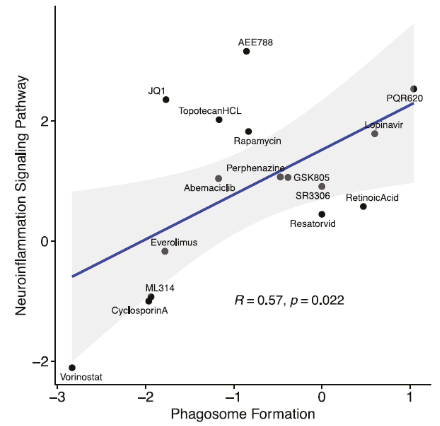
Figure 6. Scatterplot showing the relationship between phagosome formation and neuroinflammation pathways for the 16 “RNA-active” compounds exhibiting ≤50% DMSO control phagocytic index. Vorinostat is the most promising candidate, shown in the bottom left. Figure from McCrea and Batorsky et al., 2025.
This decoupling suggests that it is possible to suppress deleterious microglial activity, like excessive synaptic pruning, without triggering off-target immune responses; essential considerations for clinical translation. As vorinostat is already used as a mono or combinatorial therapy to treat cutaneous T-cell lymphoma (CTCL), has a favorable safety and tolerability profile, and has shown promising neurological results in animal models, it is a particularly attractive candidate for drug repositioning [9].
Overall, the secondary screen with MERCURIUS™ DRUG-seq resulted in a mechanistic resolution impossible to achieve with high-content imaging alone. It uncovered diverse transcriptional pathways affected by treatment, provided high-dimensional expression data necessary for deciding which therapeutic compounds to progress in preclinical pipelines, and gave insights into the precise mechanisms these drugs use to modulate microglial function.
Discussion
This study demonstrates the power of combining phenotypic high-content screening with high-throughput transcriptomic technologies to resolve the complex cellular effects and molecular mechanisms affected by small-molecule treatment.
While high-content imaging and morphological screens are perfect for triaging large compound libraries to detect the desired cellular phenotype, they miss crucial transcriptomic changes that might drive any observed cellular responses. Transcriptome-wide gene expression screening studies were previously too costly or low-throughput for large-scale screens, precluding their widespread use and limiting the amount of expression data to tens of genes (with qRT-PCR) or samples (with standard RNA-seq) at a time.
This study successfully demonstrates how MERCURIUS™ DRUG-seq effectively bridges this gap by offering cost-effective, scalable, transcriptome-wide data across large compound libraries or experimental conditions simultaneously. Compared to other multiplexed transcriptomics technologies, MERCURIUS™ DRUG-seq’s sensitivity and compatibility with 96- and 384-well workflows made it ideal for this multimodal screen and integration with exploratory high-content assays.
Importantly, the multimodal approach allowed the researchers to identify not just if a compound affects cellular phenotypes, but how likely it is to produce undesirable inflammation in microglia, allowing them to discover other off-target effects early on in therapeutic development. The results obtained by combining high-content imaging with high-throughput transcriptomics in this study could open new therapeutic avenues for treating a range of brain diseases.
Conclusion
By combining high-content imaging and MERCURIUS™ DRUG-seq, the research team:
- Efficiently screened nearly 500 CNS-penetrant compounds.
- Identified and validated 28 compounds that suppress microglial synaptic pruning.
- Distinguished promising therapeutic candidates based on both phenotypic and transcriptomic signatures.
- Highlighted compounds like vorinostat and rapamycin for potential repurposing in neurological diseases.
Integrating high-throughput imaging with scalable transcriptomics is a powerful, unbiased platform for drug discovery, particularly for complex, functional phenotypes like microglial modulation. For researchers aiming to dissect compound mechanisms of action at scale, MERCURIUS™ DRUG-seq offers the throughput, resolution, and reproducibility needed to drive discovery and development.
Contact us to discuss how combining high-content screening and MERCURIUS™ DRUG-seq can help you make the most of your next compound screen.
References
- McCrea LT, Batorsky RE, Bowen JJ, Yeh H, Thanos JM, Fu T, Perlis RH, Sheridan SD. Identifying brain-penetrant small-molecule modulators of human microglia using a cellular model of synaptic pruning. Neuropsychopharmacology. 2025 May 9:1-9.
- Neniskyte U, Gross CT. Errant gardeners: glial-cell-dependent synaptic pruning and neurodevelopmental disorders. Nature Reviews Neuroscience. 2017 Nov;18(11):658-70.
- Seal S, Trapotsi MA, Spjuth O, Singh S, Carreras-Puigvert J, Greene N, Bender A, Carpenter AE. Cell Painting: a decade of discovery and innovation in cellular imaging. Nature Methods. 2024 Dec 5:1-5.
- Alexander-Dann B, Pruteanu LL, Oerton E, Sharma N, Berindan-Neagoe I, Módos D, Bender A. Developments in toxicogenomics: understanding and predicting compound-induced toxicity from gene expression data. Molecular omics. 2018;14(4):218-36.
- Alpern D, Gardeux V, Russeil J, Mangeat B, Meireles-Filho AC, Breysse R, Hacker D, Deplancke B. BRB-seq: ultra-affordable high-throughput transcriptomics enabled by bulk RNA barcoding and sequencing. Genome biology. 2019 Dec;20:1-5.
- Sheridan SD, Horng JE, Perlis RH. Patient-derived in vitro models of microglial function and synaptic engulfment in schizophrenia. Biological psychiatry. 2022 Sep 15;92(6):470-9.
- Sellgren C, Sheridan S, Gracias J, Xuan D, Fu T, Perlis R. Patient-specific models of microglia-mediated engulfment of synapses and neural progenitors. Mol Psychiatry. 2017;22:170–177.
- Vidal-Itriago A, Radford RA, Aramideh JA, Maurel C, Scherer NM, Don EK, Lee A, Chung RS, Graeber MB, Morsch M. Microglia morphophysiological diversity and its implications for the CNS. Frontiers in immunology. 2022 Oct 19;13:997786.
- Athira K, Sadanandan P, Chakravarty S. Repurposing vorinostat for the treatment of disorders affecting brain. Neuromolecular Med. 2021;23:449–65.